What Are The Grooves In The Brain Called
listenit
Mar 28, 2025 · 7 min read
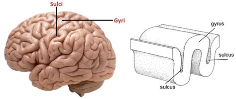
Table of Contents
- What Are The Grooves In The Brain Called
- Table of Contents
- What Are the Grooves in the Brain Called? A Deep Dive into Gyri and Sulci
- Understanding the Convoluted Cortex: Why the Grooves?
- The Significance of Surface Area: Implications for Cognitive Function
- Gyri: The Elevated Ridges of the Brain
- Key Gyri and Their Functions:
- Sulci: The Grooves That Shape the Brain
- Key Sulci and Their Significance:
- The Interplay of Gyri and Sulci: A Functional Partnership
- Developmental Aspects: Shaping the Brain's Landscape
- Functional Implications: Optimized Neural Communication
- Clinical Significance: Gyri and Sulci in Neurological Disorders
- Examples of Neurological Conditions Related to Gyri and Sulci:
- Advanced Imaging Techniques: Visualizing the Brain's Topography
- MRI and fMRI: High-Resolution Imaging
- Ongoing Research: Unraveling the Mysteries of Gyri and Sulci
- Future Directions: From Structure to Function
- Conclusion: The Importance of Understanding the Brain's Folded Landscape
- Latest Posts
- Latest Posts
- Related Post
What Are the Grooves in the Brain Called? A Deep Dive into Gyri and Sulci
The human brain, a marvel of biological engineering, isn't a smooth, featureless organ. Instead, its surface is characterized by a complex landscape of ridges and grooves, a topography crucial to its immense computational power. Understanding the names and functions of these structures is key to grasping the brain's intricate workings. This article will delve deep into the fascinating world of gyri and sulci, the ridges and grooves, respectively, that form the brain's distinctive convoluted appearance.
Understanding the Convoluted Cortex: Why the Grooves?
The brain's convoluted surface, technically known as the cerebral cortex, isn't just aesthetically interesting; it's functionally essential. This highly folded structure significantly increases the surface area of the brain within the confines of the skull. This increased surface area allows for a vast expansion of the cortical gray matter – the crucial region responsible for higher-order cognitive functions like language, reasoning, and memory.
Imagine trying to fit a large piece of fabric into a small container. Folding it neatly allows you to fit much more fabric. Similarly, the gyri and sulci allow the brain to pack a tremendous amount of neural processing power into a relatively compact space. Without this folding, the human brain would need to be significantly larger, making childbirth incredibly difficult and potentially resulting in a less efficient brain architecture.
The Significance of Surface Area: Implications for Cognitive Function
The increased surface area provided by the gyri and sulci directly translates to enhanced cognitive abilities. More cortical gray matter means more neurons and more connections (synapses) between them. This denser neural network is the basis for complex cognitive processes. This intricate arrangement is critical for the advanced capabilities that distinguish humans from other species.
Gyri: The Elevated Ridges of the Brain
Gyri (singular: gyrus) are the elevated, raised convolutions or ridges on the surface of the brain. They are separated from each other by sulci, creating the characteristic folded appearance. Individual gyri are named based on their location and often their functional specialization. Some prominent gyri include:
Key Gyri and Their Functions:
-
Precentral Gyrus: Located in the frontal lobe, it's the primary motor cortex, responsible for voluntary movement. Damage to this area can result in paralysis or weakness on the opposite side of the body. Keywords: Motor cortex, voluntary movement, paralysis.
-
Postcentral Gyrus: Situated in the parietal lobe, it's the primary somatosensory cortex, processing sensory information like touch, temperature, pain, and pressure. Lesions here can lead to sensory disturbances. Keywords: Somatosensory cortex, touch, temperature, pain, sensory disturbances.
-
Superior Temporal Gyrus: Located in the temporal lobe, this gyrus plays a crucial role in auditory processing and language comprehension. Damage can impair speech understanding (Wernicke's aphasia). Keywords: Auditory processing, language comprehension, Wernicke's aphasia.
-
Inferior Temporal Gyrus: Also in the temporal lobe, this gyrus is involved in visual object recognition and memory. Damage can affect the ability to recognize familiar faces (prosopagnosia) or objects. Keywords: Visual object recognition, memory, prosopagnosia.
-
Cingulate Gyrus: Located deep within the longitudinal fissure, it's involved in emotional regulation, cognitive control, and memory. It's part of the limbic system. Keywords: Emotional regulation, cognitive control, memory, limbic system.
-
Fusiform Gyrus: Found in the temporal and occipital lobes, it plays a critical role in facial recognition and reading. Damage can significantly impair these abilities. Keywords: Facial recognition, reading, prosopagnosia.
-
Hippocampal Gyrus: Curving around the hippocampus, it's crucial for memory consolidation and spatial navigation. This structure is tightly linked to the hippocampus's function. Keywords: Memory consolidation, spatial navigation, hippocampus.
These are just a few examples; numerous other gyri exist, each contributing to the brain's complex functional architecture. The specific functions of many gyri are still under active investigation, as neuroscience continues to unravel the intricate mysteries of the brain.
Sulci: The Grooves That Shape the Brain
Sulci (singular: sulcus) are the grooves or fissures that separate the gyri. These furrows are equally vital in shaping the brain's structure and function. Like gyri, sulci are named according to their location and are often used as landmarks to delineate different brain regions. Major sulci include:
Key Sulci and Their Significance:
-
Central Sulcus (Rolandic Sulcus): This prominent sulcus separates the frontal lobe from the parietal lobe. It's a crucial anatomical landmark easily identified on brain images. Keywords: Frontal lobe, parietal lobe, anatomical landmark.
-
Lateral Sulcus (Sylvian Fissure): A deep fissure that separates the temporal lobe from the frontal and parietal lobes. It's another easily recognizable landmark. Keywords: Temporal lobe, frontal lobe, parietal lobe, anatomical landmark.
-
Longitudinal Fissure: This is the largest sulcus, separating the two cerebral hemispheres. It's a prominent feature clearly visible on the brain's surface. Keywords: Cerebral hemispheres, brain anatomy.
-
Parieto-occipital Sulcus: This sulcus separates the parietal lobe from the occipital lobe. It's less prominent than the central or lateral sulci but still an important anatomical marker. Keywords: Parietal lobe, occipital lobe, anatomical landmark.
-
Calcarine Sulcus: Located in the occipital lobe, it houses the primary visual cortex, responsible for processing visual information. Keywords: Primary visual cortex, visual processing, occipital lobe.
These sulci, along with others less prominent, provide a framework for understanding the brain's organization. They help delineate the lobes, allowing neuroscientists to map functional areas and understand how different brain regions interact.
The Interplay of Gyri and Sulci: A Functional Partnership
Gyri and sulci aren't just passively present; they actively contribute to the brain's functionality. Their intricate interplay optimizes neural processing. The folding pattern isn't random; it's likely influenced by developmental processes and genetic factors, leading to a unique pattern in each individual brain.
Developmental Aspects: Shaping the Brain's Landscape
The formation of gyri and sulci is a complex developmental process that begins early in fetal development. Genetic factors play a significant role in determining the overall pattern of folding, but environmental factors can also influence the final morphology. Disruptions in these developmental processes can lead to various neurological conditions.
Functional Implications: Optimized Neural Communication
The close proximity of different cortical areas due to the folded architecture enhances communication and integration between brain regions. This efficient organization is essential for complex cognitive functions requiring coordination between different brain areas.
Clinical Significance: Gyri and Sulci in Neurological Disorders
Damage to specific gyri or sulci can manifest in a range of neurological symptoms, depending on the affected area. Neuroimaging techniques like MRI and fMRI allow clinicians to visualize gyri and sulci, helping to diagnose and understand various conditions.
Examples of Neurological Conditions Related to Gyri and Sulci:
-
Stroke: Damage to the blood supply to a specific gyrus can lead to focal neurological deficits, such as weakness or sensory loss.
-
Traumatic Brain Injury (TBI): Injuries impacting gyri and sulci can cause a wide range of cognitive and motor impairments.
-
Epilepsy: Abnormal electrical activity originating in a specific gyrus can trigger seizures.
-
Alzheimer's Disease: The disease affects various brain regions, including the gyri, leading to cognitive decline.
Understanding the anatomy of gyri and sulci is crucial for neurologists and neurosurgeons to accurately diagnose and treat these conditions.
Advanced Imaging Techniques: Visualizing the Brain's Topography
Modern neuroimaging techniques provide detailed visualizations of the brain's convoluted surface, allowing researchers and clinicians to study the gyri and sulci in unprecedented detail.
MRI and fMRI: High-Resolution Imaging
Magnetic resonance imaging (MRI) and functional MRI (fMRI) provide high-resolution images of the brain's structure and function. These techniques are invaluable for visualizing the gyri and sulci, allowing for precise identification of brain regions.
Ongoing Research: Unraveling the Mysteries of Gyri and Sulci
Research on gyri and sulci continues to evolve, revealing new insights into their functions and contributions to brain development and function. Advanced imaging techniques and sophisticated analytical methods are helping to unlock the mysteries of this fascinating aspect of brain anatomy.
Future Directions: From Structure to Function
Future research will likely focus on understanding the precise relationships between the structural characteristics of gyri and sulci and their specific functional roles. This includes investigating how variations in gyrification (the process of folding) relate to individual differences in cognitive abilities and susceptibility to neurological disorders. Furthermore, exploring the genetic and epigenetic factors that influence the development of these structures is crucial for understanding brain development and disease.
Conclusion: The Importance of Understanding the Brain's Folded Landscape
The gyri and sulci, the ridges and grooves of the cerebral cortex, are far more than just interesting anatomical features. Their intricate arrangement is fundamental to the brain's remarkable computational power. Understanding their morphology, function, and clinical significance is crucial for advancing our knowledge of the brain and developing effective treatments for neurological disorders. The ongoing research in this field promises to reveal even more about the complex relationship between brain structure and function, further enriching our understanding of this incredible organ.
Latest Posts
Latest Posts
-
How Elements Are Arranged In The Modern Periodic Table
Apr 01, 2025
-
Is The Square Root Of 13 Rational
Apr 01, 2025
-
How Many Protons Are In Magnesium 24
Apr 01, 2025
-
What Is The Radian Of 45 Degrees
Apr 01, 2025
-
How Many Orbitals In Each Sublevel
Apr 01, 2025
Related Post
Thank you for visiting our website which covers about What Are The Grooves In The Brain Called . We hope the information provided has been useful to you. Feel free to contact us if you have any questions or need further assistance. See you next time and don't miss to bookmark.
