Why Do Histones Bind Tightly To Dna
listenit
Mar 26, 2025 · 6 min read
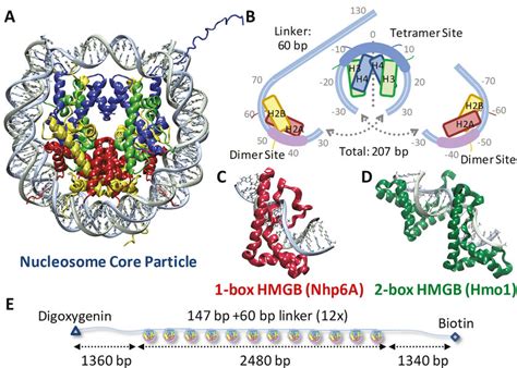
Table of Contents
Why Do Histones Bind Tightly to DNA? A Deep Dive into the Structure and Function of Nucleosomes
The human genome, a vast library of genetic information, needs to be meticulously organized to fit within the confines of a cell nucleus. This incredible feat of biological engineering is largely due to the intricate packaging of DNA around histone proteins. Histones are fundamental proteins that form the structural basis of chromatin, the complex of DNA and proteins found in eukaryotic cells. Understanding why histones bind tightly to DNA is crucial to grasping the fundamental mechanisms of gene regulation, DNA replication, and repair. This article delves into the multifaceted reasons behind this strong interaction, exploring the structural details, the forces involved, and the implications for cellular processes.
The Structural Basis of Histone-DNA Binding: The Nucleosome
The primary unit of chromatin organization is the nucleosome. A nucleosome comprises an octamer of histone proteins – two copies each of histones H2A, H2B, H3, and H4 – around which approximately 147 base pairs (bp) of DNA are wrapped in approximately 1.67 left-handed superhelical turns. This tightly wound structure drastically compacts the DNA, reducing its overall length and facilitating its organization within the nucleus.
The Histone Fold and DNA Interaction
The core histones (H2A, H2B, H3, and H4) share a conserved structural motif known as the histone fold. This motif consists of three α-helices connected by two loops, forming a compact globular domain. These histone folds mediate the interactions between the individual histones within the octamer. Importantly, the histone fold also presents surfaces that directly contact the DNA.
The DNA wraps around the histone octamer primarily through electrostatic interactions. The negatively charged phosphate backbone of DNA interacts with the positively charged amino acid residues on the histone proteins. Specifically, lysine (Lys) and arginine (Arg) residues, abundant in the histone N-terminal tails and within the histone folds, are particularly crucial for these electrostatic interactions. The high density of these positively charged amino acids creates a strong electrostatic attraction to the negatively charged DNA.
Minor Groove Interactions and Shape Complementarity
Beyond electrostatic interactions, the DNA also interacts with the histone octamer through various other forces. The DNA minor groove interacts extensively with the histone proteins. The shape complementarity between the DNA minor groove and the surfaces of the histone proteins contributes significantly to the stability of the nucleosome. Specific amino acid side chains within the histone proteins make direct contacts with the bases and sugar-phosphate backbone within the minor groove, further stabilizing the DNA-histone interaction.
Beyond Electrostatics: Other Forces Driving Histone-DNA Binding
While electrostatic interactions are dominant, several other forces contribute to the strong binding between histones and DNA:
Hydrogen Bonds
Numerous hydrogen bonds form between the DNA bases and the histone proteins. These bonds, though individually weak, collectively contribute substantially to the overall binding energy. The specific amino acid residues involved in hydrogen bonding vary depending on the DNA sequence and the specific location on the histone octamer.
Van der Waals Forces
Van der Waals forces, weak attractive forces between molecules, also play a role in histone-DNA binding. These forces contribute to the overall stability of the nucleosome structure by optimizing the packing of the DNA around the histone octamer.
Hydrophobic Interactions
Hydrophobic interactions, arising from the tendency of nonpolar molecules to cluster together in an aqueous environment, also contribute to the stability of the histone-DNA complex. Certain hydrophobic amino acid residues within the histone proteins interact with the hydrophobic parts of the DNA bases, further enhancing the binding affinity.
The Role of Histone Tails in DNA Binding and Chromatin Structure
The N-terminal tails of the core histones extend outwards from the nucleosome core particle. These tails are highly flexible and are subject to a wide range of post-translational modifications, including acetylation, methylation, phosphorylation, and ubiquitination. These modifications significantly impact the histone-DNA interaction and chromatin structure.
Post-Translational Modifications and Their Impact
- Acetylation: Acetylation of lysine residues in the histone tails neutralizes their positive charge, reducing the electrostatic attraction between the histones and DNA. This typically leads to chromatin decondensation and increased gene transcription.
- Methylation: Methylation of lysine and arginine residues can either activate or repress gene transcription depending on the specific residue and the degree of methylation. Methylation can affect the interaction of the histone tails with other proteins involved in chromatin remodeling and gene regulation.
- Phosphorylation: Phosphorylation of serine and threonine residues introduces a negative charge, altering the electrostatic interactions within the nucleosome and impacting chromatin structure. This modification often plays a critical role in processes such as DNA replication and repair.
- Ubiquitination: Ubiquitination is the attachment of ubiquitin, a small protein, to lysine residues. This modification can influence chromatin structure and gene expression through various mechanisms.
These post-translational modifications often work in concert, creating a complex regulatory landscape that governs access to DNA and controls gene expression. The interplay of different modifications, known as the "histone code," provides a nuanced mechanism for fine-tuning gene regulation.
The Importance of Histone Variants
In addition to the canonical core histones, cells also express histone variants. These variants differ slightly in amino acid sequence from the canonical histones and often have specialized roles in chromatin structure and function.
Histone Variant Functions
For instance, histone variant H2AX is involved in DNA repair, while CENP-A is a centromere-specific histone variant essential for chromosome segregation. These variants often have subtle differences in their interaction with DNA, influencing local chromatin structure and function.
Implications for Cellular Processes
The tight binding of histones to DNA is not just a structural feature; it is fundamental to several crucial cellular processes:
Gene Regulation
The accessibility of DNA to the transcriptional machinery is directly influenced by the level of compaction of chromatin. The tight wrapping of DNA around histones in the nucleosome can restrict access to genes, repressing transcription. Conversely, alterations in chromatin structure, mediated by histone modifications and remodeling complexes, can make DNA more accessible, promoting gene expression.
DNA Replication
The accurate replication of the genome requires the unwinding and duplication of DNA. This process necessitates the transient disruption of histone-DNA interactions, allowing replication machinery to access the DNA. Specialized enzymes and protein complexes facilitate the controlled disassembly and reassembly of nucleosomes during replication.
DNA Repair
Similar to DNA replication, DNA repair also requires access to the damaged DNA. The alteration of chromatin structure, often mediated by histone modifications, allows repair proteins to access the damaged region and carry out the necessary repair processes.
Chromosome Segregation
Proper chromosome segregation during cell division is essential for maintaining genomic integrity. The highly organized structure of chromatin, maintained by histone-DNA interactions, ensures that chromosomes are properly condensed and separated during mitosis and meiosis.
Conclusion
The tight binding of histones to DNA is a result of a complex interplay of various forces, including electrostatic interactions, hydrogen bonds, van der Waals forces, and hydrophobic interactions. The structural features of histones, particularly the histone fold and the N-terminal tails, are critical determinants of this interaction. Post-translational modifications of histone tails, the presence of histone variants, and the action of chromatin remodeling complexes dynamically regulate histone-DNA interactions, influencing fundamental cellular processes like gene regulation, DNA replication, repair, and chromosome segregation. Understanding the intricacies of histone-DNA binding is crucial for comprehending the fundamental workings of the eukaryotic cell and developing strategies to combat diseases associated with chromatin dysfunction. Further research continues to unravel the subtle nuances of this vital interaction, promising further insights into the regulation of gene expression and genome stability.
Latest Posts
Latest Posts
-
Why Is Density A Derived Unit
Mar 29, 2025
-
Is Density A Physical Or Chemical Change
Mar 29, 2025
-
What Plane Divides The Body Into Anterior And Posterior Parts
Mar 29, 2025
-
How Many Electron Shells Does Carbon Have
Mar 29, 2025
-
Inverse Function Of X 3 X 2
Mar 29, 2025
Related Post
Thank you for visiting our website which covers about Why Do Histones Bind Tightly To Dna . We hope the information provided has been useful to you. Feel free to contact us if you have any questions or need further assistance. See you next time and don't miss to bookmark.
