Where Is The Electron Transport Chain Located In Prokaryotes
listenit
Mar 23, 2025 · 6 min read
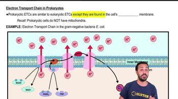
Table of Contents
Where is the Electron Transport Chain Located in Prokaryotes?
The electron transport chain (ETC), a crucial component of cellular respiration, is responsible for generating the majority of ATP (adenosine triphosphate), the cell's primary energy currency. While in eukaryotes, the ETC is neatly housed within the inner mitochondrial membrane, the location in prokaryotes is a bit more nuanced and depends on the specific organism and its metabolic strategies. This article delves into the fascinating intricacies of prokaryotic ETC location, exploring the various membrane systems involved and the implications for energy production.
Understanding the Electron Transport Chain
Before diving into prokaryotic specifics, let's briefly review the fundamental principles of the ETC. The ETC is a series of protein complexes embedded within a membrane. Electrons, harvested from the breakdown of energy-rich molecules like glucose during glycolysis and the citric acid cycle, are passed along this chain. This electron transfer releases energy, which is used to pump protons (H+) across the membrane, creating a proton gradient. This gradient then drives the synthesis of ATP via chemiosmosis, utilizing the enzyme ATP synthase.
Prokaryotic Membrane Systems: A Diverse Landscape
Unlike eukaryotic cells with their well-defined mitochondria, prokaryotes lack membrane-bound organelles. This means the ETC is not confined to a single, analogous structure. Instead, the location is dictated by the cell's plasma membrane, a highly versatile and dynamic structure. The diversity in prokaryotic membrane systems profoundly influences the ETC's localization and functionality.
1. The Plasma Membrane: The Primary Location
For many prokaryotes, the plasma membrane, also known as the cytoplasmic membrane, serves as the primary location for the ETC. This membrane separates the cytoplasm from the external environment and plays a vital role in various cellular processes, including respiration. The ETC complexes are embedded within this membrane, facilitating the proton pumping and ATP synthesis. This arrangement is particularly common in bacteria that utilize aerobic respiration.
2. Intracellular Membranes: Specialized Compartments
Some prokaryotes, particularly those engaged in phototrophy or other specialized metabolisms, have evolved intracellular membrane systems that increase the surface area available for ETC components. These membranes can form various structures, including:
-
Thylakoid membranes: In photosynthetic bacteria like cyanobacteria, these membranes are analogous to the thylakoid membranes in chloroplasts of eukaryotic plants. They are the site of photosynthesis and harbor components of the ETC involved in light-dependent reactions. The ETC here utilizes light energy to generate a proton gradient, driving ATP synthesis.
-
Mesosomes: These are invaginations of the plasma membrane, forming internal membrane folds. While their functional significance remains debated, mesosomes were once thought to be associated with respiration and the ETC in some bacteria. However, current evidence suggests that many structures previously classified as mesosomes are artifacts of fixation techniques used in electron microscopy.
-
Chromatophores: These are specialized membrane vesicles found in many photosynthetic bacteria. They contain the photosynthetic pigments and components of the ETC necessary for light harvesting and ATP production. These structures act as independent compartments optimizing the photosynthetic process.
The presence and complexity of these intracellular membranes often correlate with the organism's metabolic capabilities and its environment. For instance, bacteria thriving in low-light conditions may have more extensive internal membrane systems to maximize light capture for photosynthesis.
Variations in ETC Composition and Function
The components and precise organization of the ETC vary considerably among different prokaryotic groups. This reflects the evolutionary adaptations to diverse environments and metabolic strategies. For instance:
-
Aerobic vs. Anaerobic Respiration: Aerobic respiration, utilizing oxygen as the final electron acceptor, typically involves a longer ETC with a greater proton pumping capacity, leading to higher ATP yields. Anaerobic respiration, employing alternative electron acceptors like nitrate or sulfate, may have a shorter and less efficient ETC.
-
Phototrophy vs. Chemotrophy: Phototrophic prokaryotes utilize light energy to drive the ETC, while chemotrophic prokaryotes obtain energy from the oxidation of chemical compounds. The composition of the ETC will differ significantly between these groups, reflecting the different energy sources.
-
Gram-Positive vs. Gram-Negative Bacteria: The differences in cell wall structure between Gram-positive and Gram-negative bacteria may indirectly influence the organization of the ETC within the plasma membrane. The cell wall structure can affect the fluidity and composition of the plasma membrane, potentially impacting the distribution and function of ETC complexes.
Implications for Antibiotic Targetting
The location and composition of the ETC in prokaryotes have significant implications for the development of antibiotics. Many antibiotics target specific components of the ETC, disrupting energy production and ultimately killing the bacteria. A deeper understanding of the variations in ETC location and structure across different prokaryotic species is crucial for developing new and more effective antibiotics to combat antibiotic resistance.
The Role of Quinones and Cytochromes
The ETC is not just composed of protein complexes but also involves small, mobile electron carriers such as quinones and cytochromes. Quinones, like ubiquinone (coenzyme Q), are lipid-soluble molecules that move within the membrane, shuttling electrons between protein complexes. Cytochromes, containing heme groups, are iron-containing proteins that accept and donate electrons, participating in various steps of electron transfer. The precise arrangement and distribution of these mobile carriers within the prokaryotic membrane significantly influence the efficiency of electron transport.
Investigating ETC Location: Techniques and Challenges
Identifying the precise location of ETC components within the complex architecture of prokaryotic membranes requires sophisticated techniques. These include:
-
Electron microscopy: This technique provides high-resolution images of cellular structures, allowing visualization of membrane systems and their organization. However, it may not always resolve the precise location of individual ETC complexes.
-
Biochemical fractionation: This involves separating different cellular components, including membranes, and analyzing their protein content. This allows for the identification of ETC proteins associated with specific membrane fractions.
-
Immunogold labeling: This technique uses antibodies linked to gold particles to visualize specific proteins within the cell. By labeling ETC components, it's possible to determine their location within the membranes.
-
Proteomics: Analyzing the complete protein complement of a cell or cellular fraction helps identify all components of the ETC and can be combined with other techniques to locate them within the membrane.
Investigating the ETC in prokaryotes presents several challenges. The small size of prokaryotic cells and the dynamic nature of their membranes make it difficult to pinpoint the exact location of all ETC components. Furthermore, the diversity of prokaryotic species necessitates studying a wide range of organisms to fully understand the variations in ETC organization.
Conclusion: A Complex and Dynamic System
The location of the electron transport chain in prokaryotes is far from uniform. While the plasma membrane often serves as the primary site, various intracellular membrane systems may also house ETC components, particularly in organisms with specialized metabolisms. This diversity in location reflects the remarkable adaptability of prokaryotes to diverse environments and energy sources. Further research is needed to fully elucidate the intricacies of ETC organization in various prokaryotic groups, which will enhance our understanding of their physiology and potentially lead to the development of novel therapeutic strategies. The continuous exploration of prokaryotic membrane systems and their role in energy production remains a vibrant area of research in microbiology and cell biology.
Latest Posts
Latest Posts
-
1 8 Of A Yard Is How Many Inches
Mar 25, 2025
-
7 3 5 As An Improper Fraction
Mar 25, 2025
-
Net Ionic Equation For Hcl Naoh
Mar 25, 2025
-
What Is The Greatest Common Factor Of 75 And 30
Mar 25, 2025
-
How Many Valence Electrons Does Chloride Have
Mar 25, 2025
Related Post
Thank you for visiting our website which covers about Where Is The Electron Transport Chain Located In Prokaryotes . We hope the information provided has been useful to you. Feel free to contact us if you have any questions or need further assistance. See you next time and don't miss to bookmark.
