What Are Chromosomes Called When They Look Like X's
listenit
Mar 28, 2025 · 6 min read
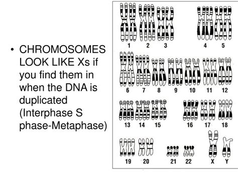
Table of Contents
What Are Chromosomes Called When They Look Like X's? A Deep Dive into Sister Chromatids and Chromosome Structure
Have you ever looked at a picture of chromosomes and wondered why some look like an "X"? That characteristic X-shape isn't just a random occurrence; it represents a crucial stage in the life cycle of a chromosome, a stage vital for cell division and the accurate transmission of genetic information. This X-shape signifies that the chromosome has duplicated, resulting in two identical copies called sister chromatids, held together at a point called the centromere. This article will explore the intricacies of chromosome structure, focusing on the "X"-shaped chromosomes and their significance in cell biology.
Understanding the Basics: What are Chromosomes?
Before delving into the X-shaped chromosomes, let's establish a foundational understanding of chromosomes themselves. Chromosomes are thread-like structures located inside the nucleus of animal and plant cells. They are made of protein and a single molecule of deoxyribonucleic acid (DNA). This DNA contains the genetic instructions for the development, functioning, growth, and reproduction of all known organisms and many viruses.
Each chromosome is composed of:
- DNA: The genetic material, carrying the genes that determine our traits. This DNA is tightly coiled and packaged around proteins called histones.
- Histones: These proteins help organize and compact the DNA, preventing it from becoming tangled and unmanageable. The combination of DNA and histones forms chromatin.
- Centromere: This is a constricted region on the chromosome, appearing as a narrow point. It plays a critical role in chromosome segregation during cell division. The centromere divides the chromosome into two arms: a short arm (p arm) and a long arm (q arm).
- Telomeres: These are protective caps located at the ends of each chromosome. They prevent the ends of chromosomes from fusing with each other and protect the genetic information from degradation.
The "X" Shape: Sister Chromatids and Chromosome Duplication
The X-shape, so often associated with chromosomes, is not a permanent state. This structure is only apparent during a specific phase of the cell cycle called M phase (Mitosis or Meiosis). This phase marks the preparation for cell division. Before the cell can divide, it must duplicate all its genetic material, including its chromosomes.
This duplication process results in two identical copies of each chromosome, joined together at the centromere. These identical copies are called sister chromatids. They are held together by a protein complex called cohesin. The cohesin ring structure keeps the sister chromatids tightly bound until they are ready to separate during cell division. This is when the iconic X-shape becomes readily visible under a microscope. The "X" therefore symbolizes the duplicated state of a chromosome, ready to be separated and distributed to daughter cells.
The Significance of Sister Chromatids
The existence of sister chromatids is crucial for accurate chromosome segregation during cell division. This ensures that each daughter cell receives a complete and identical set of chromosomes. If sister chromatids didn't exist, cell division would lead to uneven distribution of genetic material, potentially causing mutations or cell death.
Cell Division: Mitosis and Meiosis
The process of cell division is where the "X"-shaped chromosomes play their most vital role. There are two main types of cell division: mitosis and meiosis.
Mitosis: Asexual Reproduction
Mitosis is the type of cell division used for growth and repair of somatic cells (body cells). During mitosis, a single parent cell divides into two identical daughter cells. The X-shaped chromosomes are crucial in ensuring that each daughter cell receives a complete and identical copy of the genetic material.
The key stages of mitosis where the X-shaped chromosomes are particularly important are:
- Prophase: Chromosomes condense and become visible as the characteristic X-shape.
- Metaphase: Sister chromatids align along the metaphase plate (the equator of the cell).
- Anaphase: Sister chromatids separate at the centromere, moving to opposite poles of the cell. At this point, the "X" separates into two individual chromosomes.
- Telophase: The separated chromosomes reach the poles of the cell, and the nuclear membrane reforms around each set of chromosomes.
Meiosis: Sexual Reproduction
Meiosis is a specialized type of cell division that occurs in germ cells (sex cells – sperm and eggs). Unlike mitosis, meiosis results in four daughter cells, each with half the number of chromosomes as the parent cell. This reduction in chromosome number is essential for sexual reproduction, preventing the doubling of chromosome number in each generation.
The process of meiosis involves two rounds of cell division (Meiosis I and Meiosis II). Sister chromatids remain attached until Anaphase II in Meiosis II. The X-shaped chromosomes are similarly critical in ensuring accurate chromosome segregation. The process is slightly more complex in meiosis, with homologous chromosomes pairing up and undergoing recombination before separation. This recombination contributes to genetic diversity in offspring.
Chromosome Aberrations: When the "X" Goes Wrong
The precise separation of sister chromatids during cell division is essential for maintaining genomic integrity. However, sometimes errors occur, leading to chromosome abnormalities. These abnormalities can result in a variety of genetic disorders, depending on the type and extent of the error.
Some common types of chromosome aberrations involving sister chromatids include:
- Non-disjunction: Failure of sister chromatids to separate properly during anaphase, resulting in daughter cells with an abnormal number of chromosomes (aneuploidy). Examples include Down syndrome (trisomy 21), Turner syndrome (monosomy X), and Klinefelter syndrome (XXY).
- Chromatid breakage: Damage to one or both sister chromatids can lead to deletions, duplications, or translocations of genetic material. These changes can have significant consequences, depending on the genes affected.
- Sister chromatid exchange: This involves the exchange of genetic material between sister chromatids. While sometimes a normal part of cell processes, excessive sister chromatid exchange can be indicative of DNA damage or genomic instability.
Beyond the X: Other Chromosome Configurations
While the X-shape is the most commonly recognized form of a duplicated chromosome, it's important to note that the appearance of chromosomes can vary depending on the location of the centromere. The centromere's position determines the relative lengths of the p and q arms. Different centromere positions lead to different chromosome shapes:
- Metacentric: The centromere is located in the middle, resulting in two nearly equal-length arms. This results in a nearly perfect "X".
- Submetacentric: The centromere is slightly off-center, resulting in one arm being slightly longer than the other. The "X" appears slightly asymmetrical.
- Acrocentric: The centromere is located near one end, resulting in one very short arm (p arm) and one very long arm (q arm). The chromosome resembles a rod rather than an "X".
- Telocentric: The centromere is located at the end of the chromosome. Only one arm is visible. Telocentric chromosomes are relatively rare in humans.
Conclusion: The X-Shaped Chromosome – A Symbol of Genetic Fidelity
The characteristic X-shape of chromosomes is not merely a visual curiosity but a fundamental aspect of cell biology. The duplicated chromosomes, represented by the X shape and composed of two identical sister chromatids, are essential for the accurate transmission of genetic information during cell division. The precise separation of these chromatids during mitosis and meiosis ensures that daughter cells inherit the correct number and type of chromosomes, maintaining genomic stability and preventing genetic disorders. Understanding the structure and behavior of these X-shaped chromosomes is crucial for comprehending the complexities of life and the processes that govern inheritance and evolution. While deviations from the "X" exist and can have profound consequences, the structure ultimately serves as a powerful visual representation of the carefully orchestrated process of cell division and genetic continuity.
Latest Posts
Latest Posts
-
What Is The Fraction For 0 09
Mar 31, 2025
-
How To Find An Objects Acceleration
Mar 31, 2025
-
How Do Igneous Rocks Form Into Sedimentary Rocks
Mar 31, 2025
-
What Is The Sum Of A Heptagons Interior Angles
Mar 31, 2025
-
What Is The Inverse Of X 3
Mar 31, 2025
Related Post
Thank you for visiting our website which covers about What Are Chromosomes Called When They Look Like X's . We hope the information provided has been useful to you. Feel free to contact us if you have any questions or need further assistance. See you next time and don't miss to bookmark.
