The Head Of The Femur Articulates With The
listenit
Mar 18, 2025 · 6 min read
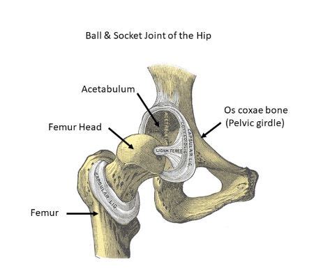
Table of Contents
The Head of the Femur Articulates With: A Deep Dive into the Hip Joint
The human hip joint, a marvel of biological engineering, is responsible for a wide range of movements, from the subtle adjustments in posture to the powerful strides of a runner. At the heart of this remarkable articulation lies the connection between the head of the femur (thigh bone) and its receiving socket, the acetabulum of the pelvis. Understanding this articulation – precisely what the head of the femur articulates with – is crucial to grasping the mechanics of locomotion, understanding hip pathologies, and appreciating the complexity of the human musculoskeletal system.
The Acetabulum: The Hip's Receiving Socket
The head of the femur doesn't articulate with a flat surface; rather, it fits snugly into a cup-shaped socket called the acetabulum. Located on the lateral aspect of the hip bone (os coxae), the acetabulum is formed by the fusion of three bones during development: the ilium, ischium, and pubis. This fusion creates a deep, secure socket that is ideally suited to receive the femoral head.
Acetabular Anatomy: A Closer Look
The acetabulum isn't simply a smooth, hemispherical cup. Its intricate anatomy contributes significantly to the stability and range of motion of the hip joint:
-
Acetabular Labrum: This fibrocartilaginous ring enhances the depth of the acetabulum, providing increased stability and improving the congruency of the joint. It acts as a shock absorber and helps to maintain the negative intra-articular pressure within the joint, crucial for maintaining stability. Tears in the labrum are a common source of hip pain.
-
Acetabular Fossa: A non-articular area at the bottom of the acetabulum. It's filled with fatty tissue and is not involved in the direct articulation with the femoral head.
-
Acetabular Facet: The articular surface of the acetabulum, covered with hyaline cartilage, providing a smooth, low-friction surface for articulation with the femoral head. This cartilage is crucial for protecting the bone and allowing for smooth, effortless movement.
-
Transverse Acetabular Ligament: This ligament spans the acetabular notch, a gap in the inferior aspect of the acetabulum, helping to complete the socket and contribute to its overall stability.
The Femoral Head: The Ball in the Socket
The femoral head, a smooth, rounded prominence at the proximal end of the femur, is the counterpart to the acetabulum. Approximately two-thirds of a sphere, it perfectly complements the concave shape of the acetabulum. This spherical shape allows for a wide range of motion in multiple planes.
Femoral Head Anatomy: Key Features
Several anatomical features of the femoral head are essential for its function within the hip joint:
-
Fovea Capitis: A small pit located centrally on the femoral head. This serves as the attachment point for the ligamentum teres, a small ligament that contributes to the hip joint's blood supply.
-
Articular Cartilage: Similar to the acetabulum, the femoral head is covered with hyaline cartilage, providing a smooth articulating surface and minimizing friction. Degeneration of this cartilage leads to osteoarthritis, a common cause of hip pain and disability.
-
Neck of the Femur: The neck connects the femoral head to the shaft of the femur, acting as a lever arm for muscle forces. Its angle and length influence the overall biomechanics of the hip. Fractures of the femoral neck are a common injury, especially in the elderly.
The Articulation: A Dynamic Interaction
The articulation between the femoral head and the acetabulum is a remarkable example of a ball-and-socket joint. This type of joint allows for a wide range of motion in three planes: flexion and extension (bending forward and backward), abduction and adduction (moving the leg away from and toward the midline of the body), and medial and lateral rotation (rotating the leg inward and outward).
Ligaments: The Hip's Stabilizers
Several crucial ligaments contribute significantly to the stability of the hip joint, preventing dislocation and guiding movement:
-
Iliofemoral Ligament: Often referred to as the "Y-ligament," it's the strongest ligament in the body, significantly contributing to hip joint stability, particularly during standing.
-
Pubofemoral Ligament: This ligament reinforces the anterior aspect of the hip joint, providing stability and limiting excessive abduction and extension.
-
Ischiofemoral Ligament: Located posteriorly, this ligament helps to limit internal rotation and provides support to the hip joint.
-
Ligamentum Teres: This intracapsular ligament connects the fovea capitis of the femoral head to the acetabulum. While it offers minimal direct support, its primary function is to carry blood vessels to the femoral head, contributing to its nourishment.
Muscles: The Drivers of Movement
Numerous muscles surround the hip joint, allowing for a wide array of coordinated movements. These muscles, originating from the pelvis and attaching to the femur, act as powerful actuators, driving the articulation between the femoral head and acetabulum.
-
Gluteal Muscles: The gluteus maximus, medius, and minimus muscles are crucial for hip extension, abduction, and external rotation.
-
Hip Flexors: Muscles like the iliopsoas, rectus femoris, and sartorius are involved in hip flexion (bringing the leg towards the torso).
-
Hip Adductors: These muscles, including the adductor longus, brevis, and magnus, draw the leg towards the midline of the body.
-
Hip Rotators: A complex group of muscles are involved in internal and external rotation, including the piriformis, obturator internus and externus, and quadratus femoris.
Clinical Significance: Understanding Hip Pathology
A thorough understanding of the articulation between the femoral head and the acetabulum is paramount in the diagnosis and treatment of numerous hip pathologies. Conditions affecting this articulation can lead to significant pain, disability, and reduced quality of life.
Common Hip Conditions:
-
Osteoarthritis: This degenerative joint disease is characterized by the breakdown of articular cartilage, leading to pain, stiffness, and limited range of motion.
-
Hip Impingement (Femoroacetabular Impingement or FAI): This condition involves abnormal contact between the femoral head and the acetabulum, leading to cartilage damage and pain.
-
Hip Labral Tears: Tears in the acetabular labrum can cause pain, clicking, and instability in the hip joint.
-
Hip Dislocation: A serious injury involving the complete separation of the femoral head from the acetabulum.
-
Avascular Necrosis (Osteonecrosis): Disruption of the blood supply to the femoral head can lead to bone death and collapse.
-
Rheumatoid Arthritis: This autoimmune disease can affect the hip joint, causing inflammation, pain, and joint destruction.
Conclusion: A Complex and Vital Articulation
The articulation between the head of the femur and the acetabulum is far more than just a simple "ball-and-socket" joint; it's a highly complex and intricately designed structure that allows for a wide range of movement while providing significant stability. Understanding its anatomy, biomechanics, and the pathologies that can affect it is crucial for healthcare professionals and anyone interested in human movement and musculoskeletal health. From the subtle movements of daily life to the powerful strides of an athlete, the hip joint, and the perfect articulation of its components, remains a testament to the marvel of biological engineering. Continued research into the intricacies of this joint will undoubtedly lead to improved diagnostics, treatments, and preventative strategies for a wide range of conditions that affect millions worldwide.
Latest Posts
Latest Posts
-
What Is 4 To The Power Of 5
Mar 18, 2025
-
Chromium Iii Oxide Why Is It Cr2o3 And Not Cro3
Mar 18, 2025
-
The Light Dependent Reactions Occur In The Stroma Of The Chloroplast
Mar 18, 2025
-
Why Are Alkyl Groups Electron Donating
Mar 18, 2025
-
What Is The Longest Phase In Mitosis
Mar 18, 2025
Related Post
Thank you for visiting our website which covers about The Head Of The Femur Articulates With The . We hope the information provided has been useful to you. Feel free to contact us if you have any questions or need further assistance. See you next time and don't miss to bookmark.
