The Plasma Membrane Of A Muscle Cell Is Called The
listenit
Mar 28, 2025 · 6 min read
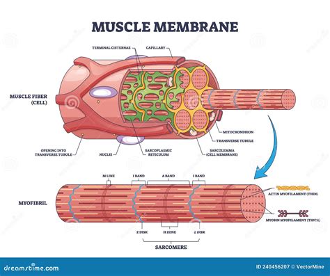
Table of Contents
The Plasma Membrane of a Muscle Cell is Called the Sarcolemma: A Deep Dive into its Structure, Function, and Clinical Significance
The plasma membrane, the selectively permeable barrier surrounding all cells, plays a vital role in maintaining cellular integrity and facilitating communication with the external environment. In muscle cells, this membrane has a specialized name: the sarcolemma. Understanding the sarcolemma's unique structure and function is crucial to comprehending muscle contraction, excitation-contraction coupling, and various muscle-related diseases. This article will delve into the intricacies of the sarcolemma, exploring its composition, specialized features, and its clinical relevance.
The Sarcolemma: More Than Just a Membrane
While fundamentally similar to the plasma membranes of other cells, the sarcolemma possesses several unique adaptations that reflect its specialized role in muscle physiology. These adaptations are crucial for efficient excitation-contraction coupling, the process that links electrical stimulation to mechanical contraction.
Structural Components of the Sarcolemma:
The sarcolemma is a complex structure comprised of several key components:
-
Phospholipid Bilayer: Like all plasma membranes, the sarcolemma’s foundation is a phospholipid bilayer. This double layer of phospholipid molecules forms a hydrophobic barrier that separates the intracellular and extracellular environments. This barrier is crucial for maintaining the cell's internal environment and regulating the passage of ions and molecules.
-
Membrane Proteins: Embedded within the phospholipid bilayer are numerous proteins, crucial for the sarcolemma's diverse functions. These proteins can be broadly classified into:
- Ion Channels: These integral membrane proteins form pores that allow specific ions (e.g., Na+, K+, Ca2+) to passively cross the membrane. Their selective permeability is essential for generating and propagating action potentials. Voltage-gated sodium channels, for instance, are critical for the rapid depolarization phase of the action potential, while voltage-gated potassium channels are responsible for repolarization.
- Ion Pumps: These proteins actively transport ions against their concentration gradients, maintaining the ionic balance crucial for muscle excitability. The sodium-potassium pump (Na+/K+ ATPase) is a prime example, expelling sodium ions and importing potassium ions, both essential for establishing the resting membrane potential. The sarcoplasmic reticulum Ca2+-ATPase (SERCA) pump is also crucial, pumping calcium ions back into the sarcoplasmic reticulum after muscle contraction.
- Receptors: These proteins bind to specific ligands (e.g., neurotransmitters, hormones) triggering intracellular signaling cascades that can modulate muscle function. Nicotinic acetylcholine receptors at the neuromuscular junction are vital for initiating muscle contraction.
- Structural Proteins: These proteins provide structural integrity and support to the sarcolemma. Examples include dystrophin, a crucial protein linking the sarcolemma to the extracellular matrix and the cytoskeleton, and integrins, which mediate cell-matrix interactions. Defects in these proteins can lead to muscular dystrophies.
-
Glycocalyx: The outer surface of the sarcolemma is coated with a glycocalyx, a layer of glycoproteins and glycolipids. This layer plays a role in cell adhesion, recognition, and protection.
Specialized Features of the Sarcolemma:
Beyond its basic structure, the sarcolemma exhibits several specialized features crucial for muscle function:
-
Transverse Tubules (T-tubules): These invaginations of the sarcolemma extend deep into the muscle fiber, forming a network that encircles the myofibrils. T-tubules play a critical role in excitation-contraction coupling by rapidly conducting action potentials from the surface of the muscle fiber to the interior, ensuring synchronous contraction of the sarcomeres.
-
Junctional Folds: At the neuromuscular junction, the sarcolemma forms junctional folds, increasing the surface area available for acetylcholine receptors. This amplification ensures efficient neurotransmission and robust muscle activation.
-
Costameres: These protein complexes link the sarcolemma to the underlying cytoskeleton and extracellular matrix, providing structural support and enabling force transmission during muscle contraction. Costameres play a critical role in maintaining the integrity of the muscle fiber and preventing damage during repeated contractions.
The Sarcolemma's Role in Excitation-Contraction Coupling:
The sarcolemma is the central player in excitation-contraction coupling, the intricate process that translates an electrical signal (action potential) into a mechanical response (muscle contraction). This process unfolds as follows:
-
Neuromuscular Transmission: An action potential arrives at the motor neuron's axon terminal, triggering the release of acetylcholine into the synaptic cleft.
-
Sarcolemma Depolarization: Acetylcholine binds to nicotinic acetylcholine receptors on the sarcolemma, causing depolarization of the muscle fiber membrane. This depolarization spreads rapidly along the sarcolemma and into the T-tubules.
-
Calcium Release: The depolarization wave activates voltage-sensitive dihydropyridine receptors (DHPRs) in the T-tubules. These receptors are mechanically coupled to ryanodine receptors (RyRs) on the sarcoplasmic reticulum (SR), the intracellular calcium store. Activation of DHPRs triggers the opening of RyRs, releasing Ca2+ from the SR into the cytoplasm.
-
Muscle Contraction: The increased cytosolic Ca2+ concentration binds to troponin C, initiating a series of events that lead to the sliding of actin and myosin filaments, resulting in muscle contraction.
-
Relaxation: Once the action potential ceases, Ca2+ is actively pumped back into the SR by SERCA, leading to muscle relaxation.
Clinical Significance of the Sarcolemma:
Dysfunction of the sarcolemma can lead to a range of debilitating muscle diseases. Several conditions directly affect the sarcolemma's structure and function:
-
Muscular Dystrophies: These genetic disorders are characterized by progressive muscle weakness and degeneration. Many muscular dystrophies result from mutations in genes encoding sarcolemma-associated proteins, such as dystrophin. The absence or dysfunction of these proteins compromises the sarcolemma's structural integrity, leading to muscle fiber damage and cell death. Duchenne muscular dystrophy, a severe form of the disease, is caused by mutations in the dystrophin gene.
-
Myasthenia Gravis: This autoimmune disorder affects the neuromuscular junction. Antibodies target and destroy acetylcholine receptors on the sarcolemma, impairing neuromuscular transmission and causing muscle weakness and fatigue.
-
Lambert-Eaton Myasthenic Syndrome (LEMS): In LEMS, antibodies target voltage-gated calcium channels at the presynaptic nerve terminal, reducing acetylcholine release and leading to muscle weakness.
-
Periodic Paralysis: This group of disorders is characterized by episodic attacks of muscle weakness or paralysis. These conditions can be caused by mutations affecting ion channels in the sarcolemma, disrupting the normal ionic balance and leading to impaired muscle excitability.
Future Research Directions:
Ongoing research continues to unravel the intricate details of sarcolemma structure and function. Areas of active investigation include:
-
Developing novel therapies for muscular dystrophies: Researchers are actively exploring gene therapy, cell-based therapies, and pharmacological interventions to treat these devastating diseases. Understanding the precise roles of sarcolemma-associated proteins is crucial for developing effective therapeutic strategies.
-
Investigating the role of the sarcolemma in muscle aging: Age-related changes in sarcolemma composition and function contribute to age-related muscle loss (sarcopenia). Understanding these changes could lead to the development of interventions to maintain muscle health in older adults.
-
Exploring the role of the sarcolemma in muscle regeneration: The sarcolemma plays a role in the process of muscle repair and regeneration after injury. Further research into these mechanisms could lead to improved therapies for muscle injuries.
Conclusion:
The sarcolemma, the plasma membrane of a muscle cell, is far more than just a simple boundary. Its complex structure, encompassing a phospholipid bilayer, a diverse array of membrane proteins, and specialized features such as T-tubules and costameres, is essential for muscle function. Its crucial role in excitation-contraction coupling, the process that translates electrical signals into muscle contraction, highlights its importance. Furthermore, understanding the sarcolemma's structure and function is crucial for comprehending a wide range of muscle diseases, paving the way for the development of novel therapeutic strategies. Future research promises to further elucidate the complexities of this critical membrane, leading to advancements in the treatment and prevention of muscle disorders.
Latest Posts
Latest Posts
-
Starting Substances In A Chemical Reaction
Mar 31, 2025
-
What Is 7 Percent As A Decimal
Mar 31, 2025
-
Choose The Graph Of Y X2 4x 5
Mar 31, 2025
-
Which Color Has The Highest Energy
Mar 31, 2025
-
How Do You Simplify Cube Roots
Mar 31, 2025
Related Post
Thank you for visiting our website which covers about The Plasma Membrane Of A Muscle Cell Is Called The . We hope the information provided has been useful to you. Feel free to contact us if you have any questions or need further assistance. See you next time and don't miss to bookmark.
