The Lungs Are Lateral To The Heart
listenit
Mar 14, 2025 · 6 min read
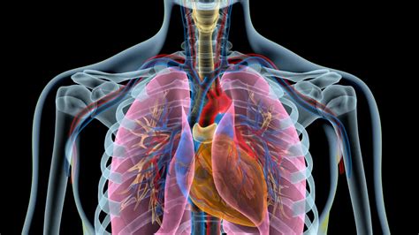
Table of Contents
The Lungs are Lateral to the Heart: Understanding Thoracic Anatomy and its Clinical Significance
The statement "the lungs are lateral to the heart" is a fundamental concept in human anatomy. Understanding this spatial relationship is crucial not only for medical professionals but also for anyone seeking a deeper understanding of the human body. This article will delve into the precise anatomical positioning of the lungs and heart, exploring the implications of their arrangement, common pathologies affecting this region, and the diagnostic tools used to assess its health.
The Thoracic Cavity: A Complex Arrangement
The heart and lungs reside within the thoracic cavity, a bony cage formed by the ribs, sternum, and thoracic vertebrae. This cavity is not a simple, open space; rather, it's intricately structured, with various membranes and compartments separating and supporting its vital organs. The pleura, a double-layered serous membrane, encloses each lung, creating a pleural cavity filled with a small amount of lubricating fluid. This fluid minimizes friction during respiration. The pericardium, another serous membrane, similarly surrounds the heart, creating a pericardial cavity with its own lubricating fluid. These serous membranes are essential for the proper functioning of both organs.
Defining Lateral and Medial Positions
Before diving into the specific location of the lungs relative to the heart, it's important to understand the anatomical directional terms. Lateral refers to structures situated away from the midline of the body, towards the sides. Medial, conversely, refers to structures closer to the midline. Anterior indicates structures towards the front, while posterior denotes structures towards the back. Superior indicates a higher position, and inferior a lower position. Using these terms precisely is vital for clear communication in anatomy and medicine.
The Heart's Central Position
The heart, a muscular pump responsible for circulating blood throughout the body, is situated medially within the thoracic cavity. It's not perfectly centered, however. Approximately two-thirds of the heart lies to the left of the midline, while the remaining third extends to the right. This slight leftward displacement is a significant anatomical feature. The heart's position is further defined by its relation to other structures; it sits anterior to the vertebral column and posterior to the sternum.
The Lungs: Flanking the Heart
Now, we can address the central theme: the lungs are lateral to the heart. This means they are situated on either side of the heart, flanking it. The right lung is slightly larger than the left, largely due to the space occupied by the heart on the left side of the chest cavity. The lungs are cone-shaped organs, extending from the clavicles superiorly to the diaphragm inferiorly. Their expansive nature allows for efficient gas exchange, vital for respiration.
Lobes and Fissures
Both lungs are further subdivided into lobes. The right lung has three lobes (superior, middle, and inferior), separated by fissures. The left lung, smaller due to the cardiac notch accommodating the heart, has two lobes (superior and inferior), separated by a single fissure. These lobes are not simply arbitrary divisions; they represent functional units within the lung. Understanding their structure is important for interpreting imaging studies and localizing lung pathology.
Clinical Significance of the Spatial Relationship
The spatial arrangement of the lungs and heart has profound clinical implications. Many diseases and conditions affecting one organ can impact the other due to their proximity and shared anatomical space. For example:
1. Cardiac Tamponade:
A life-threatening condition where fluid accumulates in the pericardial cavity, compressing the heart and hindering its ability to pump efficiently. This compression can also affect the lungs, leading to shortness of breath and reduced lung capacity. The close relationship between the heart and lungs makes early diagnosis and treatment crucial.
2. Pneumonia:
Infection of the lung tissue can spread to adjacent structures if left untreated. Severe pneumonia can affect the heart's function and potentially lead to complications. The lateral position of the lungs makes them susceptible to infections, highlighting the importance of preventative measures like vaccination.
3. Lung Cancer:
Tumors arising in the lungs can directly compress the heart or its major blood vessels, leading to various cardiovascular complications. This interaction underscores the importance of early detection and treatment of lung cancer.
4. Pulmonary Embolism:
A blood clot traveling to the lungs can block blood flow, causing respiratory distress and potentially impacting the heart. This highlights the interconnected nature of the cardiovascular and respiratory systems. The spatial relationship between the heart and lungs is directly involved in the pathogenesis of pulmonary embolism.
5. Trauma:
Chest injuries can affect both the heart and lungs simultaneously. Blunt force trauma, for example, can cause lung contusions (bruising) and cardiac contusions (bruising), creating a complex clinical picture. The proximity of the organs necessitates a comprehensive assessment in cases of trauma.
Diagnostic Imaging Techniques
Several diagnostic imaging techniques are employed to visualize the heart and lungs and assess their relative positions and health. These include:
1. Chest X-ray:
A readily available and relatively inexpensive method for visualizing the lungs, heart, and surrounding structures. It provides a two-dimensional image, allowing for the assessment of lung volume, heart size, and the presence of any abnormalities.
2. Computed Tomography (CT) Scan:
Offers a detailed three-dimensional reconstruction of the thoracic cavity, providing a much more precise visualization of the heart and lungs. It's particularly useful for identifying subtle abnormalities, such as small nodules in the lungs or subtle cardiac anomalies.
3. Magnetic Resonance Imaging (MRI):
Another powerful imaging technique, particularly useful for visualizing soft tissues. MRI provides detailed images of the heart and its structures, allowing for assessment of cardiac function and detection of abnormalities. It can also be used to visualize the lungs and surrounding structures, though it is less frequently used for routine lung assessments compared to CT scans.
4. Echocardiography:
A non-invasive ultrasound technique used specifically to visualize the heart's structure and function. It provides real-time images of the heart's chambers, valves, and blood flow, helping diagnose various cardiac conditions.
Conclusion
The statement, "the lungs are lateral to the heart," is more than just an anatomical fact; it's a cornerstone for understanding the intricate relationship between these two vital organs. Their close proximity and shared anatomical space have profound clinical implications, influencing the presentation, diagnosis, and treatment of numerous diseases. The use of various diagnostic imaging techniques allows for detailed visualization and assessment of the heart and lungs, ensuring appropriate clinical management. A thorough understanding of thoracic anatomy is essential for medical professionals and anyone interested in the workings of the human body. This intricate interplay emphasizes the interconnectedness of physiological systems and highlights the importance of a holistic approach to healthcare. Future research continues to deepen our understanding of the interactions between the heart and lungs, leading to advancements in diagnosis and treatment strategies.
Latest Posts
Latest Posts
-
Least Common Multiple For 3 4 5
Mar 14, 2025
-
What Happens To The Electrons In Ionic Bonding
Mar 14, 2025
-
X Square Root Of X 6
Mar 14, 2025
-
What Is The Square Root Of 33
Mar 14, 2025
-
What Is The Square Root Of 136
Mar 14, 2025
Related Post
Thank you for visiting our website which covers about The Lungs Are Lateral To The Heart . We hope the information provided has been useful to you. Feel free to contact us if you have any questions or need further assistance. See you next time and don't miss to bookmark.
