Name The Muscle That Subdivides The Ventral Body Cavity
listenit
Mar 19, 2025 · 6 min read
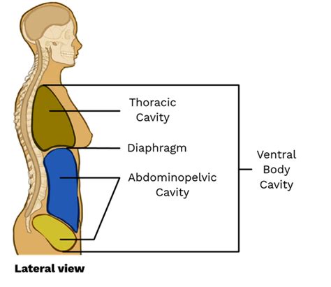
Table of Contents
The Diaphragm: The Muscle That Subdivides the Ventral Body Cavity
The human body is a marvel of intricate design, with various systems working in harmony to maintain life. One crucial aspect of this design is the organization of internal organs within distinct cavities. The ventral body cavity, a large space encompassing the chest and abdomen, plays a vital role in protecting and housing essential organs. A key structure that defines and separates this cavity is the diaphragm, a dome-shaped sheet of skeletal muscle and connective tissue. This article will delve into the anatomy, function, and clinical significance of the diaphragm, emphasizing its critical role in subdividing the ventral body cavity.
Anatomy of the Diaphragm: A Detailed Look
The diaphragm's unique structure is intrinsically linked to its function. It's not simply a flat muscle; its dome-like shape is crucial for its respiratory actions. Let's examine its key anatomical features:
Muscular Components:
- Sternal Part: This relatively small portion originates from the posterior surface of the xiphoid process of the sternum.
- Costal Part: The largest portion, originating from the inner surfaces of the lower six ribs and their corresponding costal cartilages. These fibers interdigitate with the fibers of the transversus abdominis muscle.
- Lumbar Part: Originating from two crura (tendinous structures) that arise from the lumbar vertebrae (L1-L3) and the anterior longitudinal ligament of the vertebral column. The right crus is typically longer and thicker than the left. The medial arcuate ligament and lateral arcuate ligament also contribute to this part's origin. These ligaments bridge the gap between the transverse processes of the lumbar vertebrae and the quadratus lumborum muscle.
Central Tendon:
All three muscular components converge on a central tendon, a thin, aponeurotic structure that forms the central part of the diaphragm. This tendon is composed of dense connective tissue, providing a strong, yet flexible attachment point for the muscular fibers. Its unique structure allows for efficient force transmission during contraction and relaxation.
Openings in the Diaphragm:
The diaphragm isn't a completely sealed structure. Several important openings allow the passage of structures between the thoracic and abdominal cavities:
- Aortic Hiatus: Located at the level of T12, this opening allows passage for the aorta, thoracic duct, and azygos vein. It's posterior to the diaphragm's central tendon, situated between the two crura.
- Esophageal Hiatus: Located at the level of T10, this opening transmits the esophagus and the anterior and posterior vagal trunks. It's also situated posterior to the central tendon. The esophageal hiatus allows the passage of food to the stomach without compromising the integrity of the diaphragm.
- Caval Foramen (Vena Cava Foramen): Located at the level of T8, this opening is the largest of the three and allows passage for the inferior vena cava. This opening is unique in that it's located within the central tendon itself. This location is crucial as the inferior vena cava's passage must not be impeded by the diaphragm's movement.
Functional Role of the Diaphragm: More Than Just Breathing
The diaphragm's primary function is undeniably crucial in respiration, but its influence extends beyond that, affecting other vital bodily processes.
Respiration:
During inspiration, the diaphragm contracts, causing it to flatten and descend. This increases the volume of the thoracic cavity, reducing the intrapleural pressure and drawing air into the lungs. During expiration, the diaphragm relaxes, returning to its dome shape, decreasing the thoracic volume, and expelling air from the lungs. This rhythmic contraction and relaxation is essential for normal breathing.
Support of Abdominal Viscera:
The diaphragm provides significant support to the abdominal organs, acting as a dynamic floor for the thoracic cavity and a roof for the abdominal cavity. This support is especially important during activities involving increased intra-abdominal pressure, such as coughing, sneezing, and defecation. Its role in maintaining abdominal pressure is vital for proper functioning of the gastrointestinal tract.
Coughing and Sneezing:
The diaphragm plays a crucial role in these forceful expulsive actions. Its forceful contraction contributes to the generation of the high pressures needed to expel irritants from the respiratory tract.
Vomiting and Defecation:
The diaphragm's contraction assists in increasing intra-abdominal pressure, aiding in the expulsion of vomit and feces. Its coordinated action with other abdominal muscles is key to the success of these processes.
Blood Flow Regulation:
The diaphragm's movement influences venous return to the heart. Its descent during inspiration increases abdominal pressure, promoting venous blood flow back to the heart.
Clinical Significance of the Diaphragm: Conditions and Considerations
Understanding the diaphragm's function and anatomy is crucial in diagnosing and treating various conditions. Several clinical aspects highlight its significance:
Diaphragmatic Hernia:
This condition occurs when a portion of an abdominal organ protrudes through the diaphragm into the thoracic cavity. This can cause respiratory distress, digestive issues, and cardiac complications depending on the size and location of the hernia.
Diaphragmatic Eventration:
This is a condition where the diaphragm is abnormally high and flattened, reducing its ability to function effectively. It can lead to respiratory insufficiency and other complications.
Diaphragmatic Paralysis:
Paralysis of the diaphragm can occur due to nerve damage or other neurological conditions. This results in impaired breathing and can be life-threatening.
Hiatal Hernia:
This is a specific type of hernia where a portion of the stomach protrudes through the esophageal hiatus. Symptoms can range from mild discomfort to severe heartburn and gastroesophageal reflux disease (GERD).
Diaphragmatic Rupture:
This is a serious injury that can occur due to trauma, often in car accidents or other high-impact events. It requires immediate medical attention.
Pleuritis (Pleurisy):
Inflammation of the pleura, the membranes surrounding the lungs, can also affect the diaphragm. The pain associated with pleuritis can often be referred to the shoulder and neck, due to the shared innervation of the diaphragm and these regions.
Respiratory Distress Syndrome:
In newborns, underdeveloped diaphragms can contribute to respiratory distress syndrome, a condition requiring intensive care.
Diagnostic Techniques for Diaphragmatic Issues
Several diagnostic techniques are used to assess diaphragm function and identify potential problems:
- Chest X-ray: A common initial imaging technique to visualize the diaphragm and detect abnormalities such as hernias or eventration.
- Computed Tomography (CT) Scan: Provides a more detailed three-dimensional image of the diaphragm and surrounding structures.
- Magnetic Resonance Imaging (MRI): Used to obtain high-resolution images and assess soft tissue structures in detail.
- Electromyography (EMG): Measures the electrical activity of the diaphragm to assess its neuromuscular function.
- Phrenic Nerve Conduction Study: Assesses the function of the phrenic nerve, which controls the diaphragm.
Conclusion: The Diaphragm's Unsung Importance
The diaphragm, a seemingly simple muscle, plays a multifaceted and vital role in maintaining the body's homeostasis. Its function in respiration, support of abdominal organs, and involvement in various other physiological processes underscores its critical importance. Understanding its anatomy, function, and clinical significance is essential for healthcare professionals in diagnosing and managing a wide range of conditions. Its pivotal role in subdividing the ventral body cavity allows for the efficient and protected housing of crucial organs, highlighting its unsung importance in the complex machinery of the human body. Further research into the diaphragm's complexities continues to reveal its intricate influence on overall health and wellbeing. From its contribution to respiratory function to its role in maintaining abdominal pressure, the diaphragm's impact is far-reaching and vital to human survival.
Latest Posts
Latest Posts
-
4 2 5 As An Improper Fraction
Mar 19, 2025
-
How To Find Axis Of Symmetry Parabola
Mar 19, 2025
-
Least Common Factor Of 7 And 9
Mar 19, 2025
-
What Do The Arrows In The Food Chain Represent
Mar 19, 2025
-
What Is The Solution To The Following System
Mar 19, 2025
Related Post
Thank you for visiting our website which covers about Name The Muscle That Subdivides The Ventral Body Cavity . We hope the information provided has been useful to you. Feel free to contact us if you have any questions or need further assistance. See you next time and don't miss to bookmark.
