What Structure Is Produced When Protein Fibers Radiate From Centrioles
listenit
Mar 19, 2025 · 6 min read
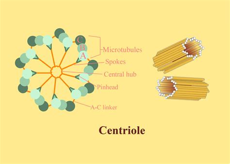
Table of Contents
What Structure is Produced When Protein Fibers Radiate from Centrioles? The Formation and Function of the Centrosome and Mitotic Spindle
The intricate dance of cell division relies on a precise orchestration of cellular components. Central to this process is the centrosome, a remarkable organelle whose structure is defined by the radiating protein fibers emanating from its centrioles. Understanding the structure produced when these fibers radiate is crucial to grasping the mechanics of cell division and the wider implications for cellular health and disease. This article will delve into the detailed structure of this complex, exploring the composition, formation, and crucial role of the centrosome and the mitotic spindle in cell division.
The Centrosome: More Than Just Two Centrioles
While often simplified as just a pair of centrioles, the centrosome is a far more complex structure. It serves as the main microtubule organizing center (MTOC) in animal cells, orchestrating the assembly and organization of microtubules—protein fibers crucial for various cellular processes, including cell division, intracellular transport, and maintaining cell shape.
Centriole Structure: A Cylindrical Masterpiece
The centrioles themselves are cylindrical structures, approximately 0.4 μm in diameter and 0.3 μm in length. They are composed of nine triplets of microtubules arranged in a cartwheel pattern. These microtubules are not simply randomly arranged; their precise organization is vital for the centrosome's function. Each triplet consists of three microtubules (A, B, and C), with the A tubule being the complete microtubule and the B and C tubules sharing some structural components with the A tubule. This intricate arrangement provides structural integrity and acts as a scaffold for the recruitment of other proteins. The precise arrangement and interactions of these microtubules are still an active area of research, with ongoing investigations exploring the role of various associated proteins.
The Pericentriolar Material (PCM): The Heart of Centrosome Activity
Surrounding the centrioles is a dense, amorphous cloud of material known as the pericentriolar material (PCM). This is where the magic happens. The PCM is a complex mixture of hundreds of proteins, including those involved in microtubule nucleation, anchoring, and regulation. This intricate protein network is responsible for:
- Microtubule Nucleation: The PCM provides the platform for the initiation of new microtubule growth. Specific proteins within the PCM, like γ-tubulin ring complexes (γ-TuRCs), act as templates for the addition of α- and β-tubulin dimers, forming the microtubule polymers.
- Microtubule Anchoring: The PCM serves as an anchoring point for the minus ends of microtubules, while the plus ends dynamically grow and shrink, exploring the cell's cytoplasm.
- Microtubule Regulation: A host of other proteins in the PCM regulate microtubule dynamics, controlling their growth, shrinkage, and stability. This dynamic regulation is essential for the accurate organization of microtubules during cell division.
The Radiating Protein Fibers: The Mitotic Spindle Takes Shape
When the cell enters mitosis, the centrosome undergoes duplication, leading to two centrosomes that migrate to opposite poles of the cell. From these centrosomes, microtubules radiate outwards, forming the mitotic spindle. This spindle is the engine of chromosome segregation, ensuring that each daughter cell receives a complete set of chromosomes.
The Three Main Types of Microtubules in the Mitotic Spindle:
The radiating microtubules in the mitotic spindle are not all the same. They can be broadly categorized into three types:
- Astral Microtubules: These microtubules radiate outwards from the centrosomes, interacting with the cell cortex, helping to position the spindle within the cell. They play a crucial role in spindle orientation and positioning.
- Kinetochore Microtubules: These microtubules connect to the kinetochores, protein structures assembled on the centromeres of chromosomes. Their dynamic instability is crucial for chromosome congression and segregation during mitosis. The attachment and detachment of kinetochore microtubules to the kinetochores is a tightly regulated process, ensuring accurate chromosome segregation.
- Interpolar Microtubules: These microtubules extend from one centrosome to the other, overlapping in the spindle midzone. They are involved in spindle elongation and are crucial for the separation of the two daughter cells during cytokinesis. The sliding movement of interpolar microtubules, driven by motor proteins, is responsible for the pushing forces that separate the poles during anaphase.
Dynamic Instability: The Driving Force of Spindle Assembly and Function
Microtubules are not static structures. They exhibit dynamic instability, a process characterized by periods of rapid growth (polymerization) followed by periods of rapid shrinkage (depolymerization). This dynamic instability is crucial for:
- Spindle Assembly: The continuous cycles of growth and shrinkage allow microtubules to explore the cell's cytoplasm, effectively searching for and attaching to kinetochores.
- Chromosome Alignment: Dynamic instability ensures that microtubules can correctly attach to kinetochores, facilitating the proper alignment of chromosomes at the metaphase plate.
- Chromosome Segregation: The regulated depolymerization of kinetochore microtubules during anaphase is crucial for pulling the sister chromatids apart, ensuring accurate chromosome segregation.
Beyond Mitosis: Centrosome Function in Other Cellular Processes
The centrosome and its radiating microtubules are not limited to cell division. They play important roles in various cellular processes, including:
- Intracellular Transport: Microtubules act as tracks for motor proteins, such as kinesins and dyneins, which transport organelles and other cellular components throughout the cell. The centrosome plays a key role in organizing these transport pathways.
- Cell Shape and Motility: The microtubule network, organized by the centrosome, contributes significantly to cell shape and motility. In some cells, microtubules extend to the cell periphery, interacting with the cell cortex and contributing to cell polarization and directed movement.
- Cilia and Flagella Formation: In ciliated and flagellated cells, the centrosome plays a crucial role in the formation of basal bodies, which serve as the anchoring points for cilia and flagella. These structures are essential for cell movement and sensory functions.
Centrosome Dysfunction and Disease
Given its central role in cell division and other essential cellular processes, it is not surprising that centrosome dysfunction is implicated in various human diseases, including:
- Cancer: Centrosome amplification, where cells have more than two centrosomes, is a common feature of many cancer types. This aberrant centrosome number can lead to chromosome instability and contribute to tumorigenesis.
- Neurodevelopmental Disorders: Disruptions in centrosome function have been linked to neurodevelopmental disorders, affecting brain development and neuronal migration.
- Genetic Diseases: Mutations in genes encoding centrosomal proteins have been associated with various genetic diseases, often characterized by developmental defects and cellular dysfunction.
Conclusion: A Complex Structure with Profound Implications
The structure produced when protein fibers radiate from centrioles—the centrosome and mitotic spindle—is far more than just a collection of protein filaments. It is a highly organized and dynamic structure, essential for cell division and numerous other cellular processes. The intricate interplay of proteins within the PCM, the dynamic instability of microtubules, and the precise organization of the mitotic spindle underscore the remarkable complexity and precision of cellular machinery. Further research continues to unravel the mysteries of centrosome biology, offering potential avenues for therapeutic interventions targeting diseases linked to centrosome dysfunction. This deeper understanding will be crucial for developing novel treatments and improving our understanding of the fundamental processes that sustain life.
Latest Posts
Latest Posts
-
How Many Unpaired Electrons Does Sulfur Have
Mar 19, 2025
-
12 Is 40 Percent Of What
Mar 19, 2025
-
What Is 1 6 In Fraction Form
Mar 19, 2025
-
Simplify X 2 1 X 2 1
Mar 19, 2025
-
From Net Earnings Of 740 Per Month
Mar 19, 2025
Related Post
Thank you for visiting our website which covers about What Structure Is Produced When Protein Fibers Radiate From Centrioles . We hope the information provided has been useful to you. Feel free to contact us if you have any questions or need further assistance. See you next time and don't miss to bookmark.
