What Muscle Subdivides The Ventral Body Cavity
listenit
Mar 28, 2025 · 6 min read
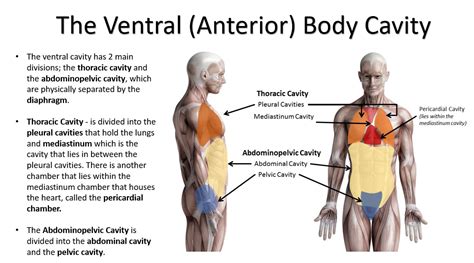
Table of Contents
What Muscle Subdivides the Ventral Body Cavity? The Diaphragm's Crucial Role
The human body is a marvel of intricate design, with various systems working in perfect harmony to maintain life. Understanding the structure and function of these systems is crucial, particularly when considering the organization of our internal organs. One key aspect of this organization is the division of the body cavity. This article delves into the crucial role of the diaphragm, the muscle responsible for subdividing the ventral body cavity into the thoracic and abdominopelvic cavities. We will explore its anatomy, function, and clinical significance in detail.
Understanding the Ventral Body Cavity
Before focusing on the diaphragm, let's establish a foundational understanding of the ventral body cavity. This large space within the body houses vital organs and is further subdivided for better protection and functional specialization. The ventral cavity is situated at the front of the body and contrasts with the dorsal cavity, which houses the brain and spinal cord. The ventral cavity is, importantly, lined by a serous membrane, a thin, double-layered membrane that secretes a lubricating fluid to minimize friction between organs and the cavity walls.
The ventral body cavity is broadly divided into two main cavities:
-
Thoracic Cavity: Located superiorly, this cavity encloses the heart, lungs, and associated structures. It's further subdivided into the pleural cavities (surrounding the lungs), the pericardial cavity (surrounding the heart), and the mediastinum (a central region containing the heart, trachea, esophagus, and major blood vessels).
-
Abdominopelvic Cavity: Located inferiorly, this cavity extends from the diaphragm to the pelvic floor. It's divided into the abdominal cavity (containing the stomach, liver, intestines, spleen, and kidneys) and the pelvic cavity (containing the bladder, rectum, and internal reproductive organs).
The Diaphragm: Anatomy and Function
The diaphragm, a dome-shaped muscle, acts as the primary anatomical structure separating the thoracic and abdominopelvic cavities. It's a crucial respiratory muscle, playing a pivotal role in breathing. Understanding its anatomy is essential to appreciate its functional significance.
Anatomical Features of the Diaphragm:
-
Shape and Position: The diaphragm's dome-like structure is positioned superiorly to the abdominal organs and inferiorly to the lungs and heart. Its central tendon, a strong aponeurosis (sheet of connective tissue), is its most prominent feature.
-
Muscle Fibers: The diaphragm's muscle fibers originate from several points: the xiphoid process (lower end of the sternum), the costal cartilages (cartilage connecting the ribs to the sternum), and the lumbar vertebrae (lower spine). These fibers converge towards the central tendon.
-
Openings: The diaphragm is not completely solid. It has several openings that allow the passage of vital structures between the thoracic and abdominopelvic cavities. These openings include the esophageal hiatus (for the esophagus), the aortic hiatus (for the aorta), and the caval opening (for the inferior vena cava).
Physiological Role of the Diaphragm in Respiration:
The diaphragm's primary function is crucial for respiration. During inspiration (inhalation), the diaphragm contracts, causing it to flatten. This downward movement increases the volume of the thoracic cavity, reducing the pressure within. This pressure difference draws air into the lungs. During expiration (exhalation), the diaphragm relaxes, returning to its dome-like shape, decreasing the thoracic cavity volume and expelling air from the lungs.
Other Functions of the Diaphragm:
Beyond its role in respiration, the diaphragm plays additional roles:
-
Support of Abdominal Viscera: The diaphragm provides support to the abdominal organs, preventing them from sagging.
-
Coughing and Sneezing: The diaphragm contributes to the forceful expulsion of air during coughing and sneezing.
-
Vomiting and Defecation: It assists in the forceful expulsion of stomach contents (vomiting) and stool (defecation).
-
Blood Circulation: Diaphragmatic movement aids venous return by increasing pressure gradients in the abdominal cavity.
Clinical Significance of the Diaphragm
The diaphragm's critical function makes it susceptible to various clinical conditions. Understanding these conditions highlights the importance of this muscle's integrity.
Diaphragmatic Hernia:
A diaphragmatic hernia is a condition where abdominal organs protrude through an opening in the diaphragm into the thoracic cavity. This can be congenital (present at birth) or acquired (resulting from trauma or injury). Symptoms can vary but may include shortness of breath, chest pain, and digestive problems.
Diaphragmatic Paralysis:
This condition occurs when the phrenic nerve (which innervates the diaphragm) is damaged, leading to paralysis of one or both sides of the diaphragm. This can result in impaired breathing, shortness of breath, and respiratory distress.
Diaphragmatic Eventration:
Diaphragmatic eventration refers to a congenital condition characterized by high-riding or flattened diaphragm. This can cause respiratory issues, particularly in infants and young children.
Hiatal Hernia:
This type of hernia involves the protrusion of the stomach through the esophageal hiatus in the diaphragm. This can lead to heartburn, acid reflux, and other gastrointestinal problems.
Respiratory Diseases:
Conditions like pneumonia, pleurisy, and pulmonary embolism can affect the diaphragm's function due to inflammation or pressure changes in the thoracic cavity.
Exploring the Interconnectedness of the Thoracic and Abdominopelvic Cavities
The diaphragm's role as a separator isn't absolute; the cavities are functionally interconnected. The shared structures and pathways highlight this crucial relationship.
Shared Structures and Pathways:
-
Esophagus: Passes through the esophageal hiatus, connecting the pharynx (throat) and stomach.
-
Aorta: The main artery of the body, passes through the aortic hiatus.
-
Inferior Vena Cava: The large vein returning blood from the lower body, passes through the caval opening.
-
Lymphatic and Nervous Systems: Lymphatic vessels and nerves traverse between the cavities, facilitating communication and coordination of bodily functions.
Functional Interdependence:
The interconnectedness isn't just anatomical; it's functional:
-
Respiratory Mechanics: Diaphragmatic movement affects both thoracic and abdominal pressures.
-
Venous Return: The respiratory pump, relying on diaphragm movement, assists venous blood return to the heart.
-
Abdominal Pressure: Intrabdominal pressure, influenced by diaphragm activity, impacts organ function and support.
-
Gastrointestinal Motility: Diaphragmatic movements can influence the motility and function of abdominal organs.
This interconnectedness underlines the significance of the diaphragm’s function. Any compromise in its integrity impacts not only respiration but also the wider physiology of both cavities.
Conclusion: The Diaphragm's Central Role
The diaphragm, with its unique anatomical structure and crucial functions, is rightfully considered the primary muscle responsible for subdividing the ventral body cavity. Its role in respiration is paramount, but its influence extends far beyond that, supporting abdominal organs and participating in various physiological processes. Understanding its anatomy, function, and clinical implications is essential for comprehending human physiology and addressing various clinical conditions. Future research is likely to further highlight the diaphragm's complexities and the nuances of its crucial role in the overall health and wellbeing of the human body. Its significance underscores the intricate relationships within the human body and the interdependent nature of its various systems.
Latest Posts
Latest Posts
-
How Do You Find Change In Velocity
Mar 31, 2025
-
What Percent Is 5 Out Of 8
Mar 31, 2025
-
Is H2s An Acid Or Base
Mar 31, 2025
-
What Is 83333 As A Fraction
Mar 31, 2025
-
Least Common Multiple 16 And 24
Mar 31, 2025
Related Post
Thank you for visiting our website which covers about What Muscle Subdivides The Ventral Body Cavity . We hope the information provided has been useful to you. Feel free to contact us if you have any questions or need further assistance. See you next time and don't miss to bookmark.
