The Joints Between Cranial Bones Of The Skull Are Called...
listenit
Apr 05, 2025 · 6 min read

Table of Contents
The Joints Between Cranial Bones of the Skull are Called Sutures: A Comprehensive Guide
The intricate architecture of the human skull is a marvel of engineering. Protecting the delicate brain, the skull isn't a single monolithic bone, but rather a complex mosaic of individual bones intricately joined together. These connections, vital for both structural integrity and the adaptability needed during birth and growth, are known as sutures. This article delves deep into the fascinating world of cranial sutures, exploring their anatomy, types, clinical significance, and the processes that affect them throughout life.
What are Cranial Sutures?
Cranial sutures are fibrous joints, specifically synarthroses, found only between the bones of the skull. Unlike the freely moving synovial joints found in the limbs, sutures are characterized by their immobility and interdigitation. The edges of the bones are intricately interwoven, creating a strong, interlocking connection that provides exceptional stability. This design minimizes movement between the bones while maintaining flexibility, particularly important during the birthing process and early childhood skull development. The fibrous connective tissue filling the space between the bony edges is primarily composed of collagen fibers, giving the suture its strength and resilience.
Types of Cranial Sutures
While all sutures are fibrous joints, they are classified based on the shape and arrangement of the interlocking bony edges:
-
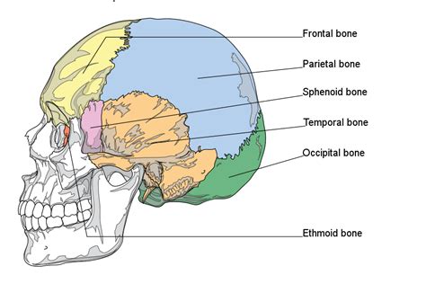
Serrate Sutures: These are the most common type of suture, characterized by interlocking, saw-tooth-like edges. The coronal suture, which separates the frontal bone from the parietal bones, and the sagittal suture, which joins the two parietal bones, are prime examples of serrate sutures. Their interlocking design provides exceptional strength and stability.
-
Squamous Sutures: In squamous sutures, the overlapping edges of the bones are beveled, creating a relatively smooth, overlapping joint. The temporoparietal suture, which connects the temporal bone to the parietal bone, is a typical example. This type of suture provides a smooth, less interlocking connection compared to serrate sutures.
-
Plane Sutures (or Butt Joints): These sutures feature relatively straight, non-overlapping edges. They are less common than serrate and squamous sutures and are found in areas where the bones meet with relatively flat surfaces. The intermaxillary suture, which connects the two maxilla bones in the upper jaw, is an example. While less intricate than other suture types, they still provide sufficient strength for their location.
-
Schindylesis: This unique type of suture involves a ridge of one bone fitting into a groove of another bone. A classic example is the articulation between the vomer and the sphenoid bone.
Major Cranial Sutures: Location and Function
Several key sutures play crucial roles in skull structure and function:
-
Coronal Suture: Runs transversely across the skull, separating the frontal bone from the two parietal bones. Its strength is vital for protecting the frontal lobes of the brain.
-
Sagittal Suture: Runs in the midsagittal plane, joining the two parietal bones. Its robustness contributes to the overall structural integrity of the skull's roof.
-
Lambdoid Suture: Shaped like the Greek letter lambda (Λ), this suture unites the occipital bone with the two parietal bones. It's critical for protecting the posterior aspects of the brain.
-
Squamous Sutures (Temporoparietal Sutures): These bilateral sutures connect the temporal bones with the parietal bones, forming the sides of the skull. Their smooth overlapping nature allows for a slightly more flexible junction.
-
Metopic Suture: This suture runs vertically down the midline of the frontal bone in infants and young children. It typically fuses completely by the age of eight, leaving a faint ridge on the frontal bone in adults.
Clinical Significance of Cranial Sutures
Cranial sutures are not merely passive structural elements; they play a significant role in various clinical conditions:
-
Craniosynostosis: This is a congenital condition where one or more sutures fuse prematurely. This can lead to abnormal skull shaping (craniofacial dysmorphism), potentially affecting brain development and causing increased intracranial pressure. Different types of craniosynostosis are named based on the affected suture (e.g., sagittal synostosis, coronal synostosis).
-
Suture Diastasis: This refers to an abnormally wide separation between the cranial bones. While sometimes asymptomatic, it can be associated with underlying skeletal abnormalities or traumatic injury.
-
Fractures: Cranial fractures can involve sutures, potentially leading to bleeding and damage to the underlying brain tissue. The location and extent of the fracture depend on the force and direction of the impact.
-
Sutural Osteoma: Benign bone tumors can form within the sutures. While usually asymptomatic, they can sometimes cause pressure symptoms or cosmetic concerns.
-
Age-Related Changes: Sutures undergo significant changes throughout life. In infants, they are relatively flexible and allow for cranial molding during birth and brain growth. As individuals age, sutures gradually ossify (fuse together), becoming increasingly rigid. This process is generally complete by adulthood, though variations exist. Premature fusion can lead to craniosynostosis, while delayed fusion can lead to conditions like craniolacuna.
The Role of Sutures in Skull Development and Growth
Cranial sutures play a pivotal role in skull development and growth, particularly during infancy and childhood. Their flexibility allows for the skull to adapt to the growing brain. The intricate interlocking of the bony edges provides strength and stability, while still allowing for limited movement and expansion. This ensures that the skull can accommodate the rapid growth of the brain during development. The timing and pattern of suture fusion are crucial for proper skull development. Premature fusion can lead to abnormal skull shape and potential complications, as discussed earlier in craniosynostosis.
Investigating Cranial Sutures: Diagnostic Techniques
Various diagnostic techniques are employed to assess the status of cranial sutures:
-
Physical Examination: A thorough physical examination, including palpation of the skull, is often the first step in assessing cranial sutures. This can reveal abnormalities such as craniosynostosis or suture diastasis.
-
Imaging Techniques: Advanced imaging techniques, such as X-rays, CT scans, and MRI, play a crucial role in visualizing cranial sutures and identifying abnormalities. These imaging modalities can provide detailed information about suture morphology, fusion status, and any associated fractures or other pathologies.
-
Genetic Testing: In some cases, genetic testing may be used to identify genetic mutations associated with craniosynostosis or other conditions affecting cranial sutures.
Sutures and the Evolution of the Skull
The study of sutures provides valuable insights into the evolutionary history of the skull. The variations in suture patterns and fusion timing across different species offer clues to evolutionary relationships and adaptive changes. The comparative anatomy of sutures across various vertebrate groups illuminates the evolution of the cranium and the development of the complex skeletal structures that protect the brain.
Conclusion: The Unsung Heroes of Skull Structure
Cranial sutures, while often overlooked, are integral components of the skull's architecture. These intricate fibrous joints provide structural strength, accommodate brain growth, and play a crucial role in the overall health and development of the cranium. Understanding their anatomy, types, clinical significance, and the processes affecting them throughout life is essential for healthcare professionals in diagnosing and managing a range of conditions affecting the skull. Further research continues to unravel the complexities of cranial suture biology, opening new avenues for improved diagnostics, treatment strategies, and a deeper understanding of human skeletal development. From their role in the birthing process to their contribution to the unique shape of our skulls, sutures represent a remarkable testament to the elegance and efficiency of biological design.
Latest Posts
Latest Posts
-
Partial Fraction Decomposition With Quadratic Factors
Apr 06, 2025
-
What Are The Factors For 29
Apr 06, 2025
-
8 Fluid Ounces To 1 Quart
Apr 06, 2025
-
What Is The Relationship Between Mutation Natural Selection And Adaptation
Apr 06, 2025
-
What Is The Main Purpose Of The Light Dependent Reactions
Apr 06, 2025
Related Post
Thank you for visiting our website which covers about The Joints Between Cranial Bones Of The Skull Are Called... . We hope the information provided has been useful to you. Feel free to contact us if you have any questions or need further assistance. See you next time and don't miss to bookmark.
