Neurotransmitter That Stimulates Skeletal Muscle Contraction
listenit
Mar 26, 2025 · 6 min read
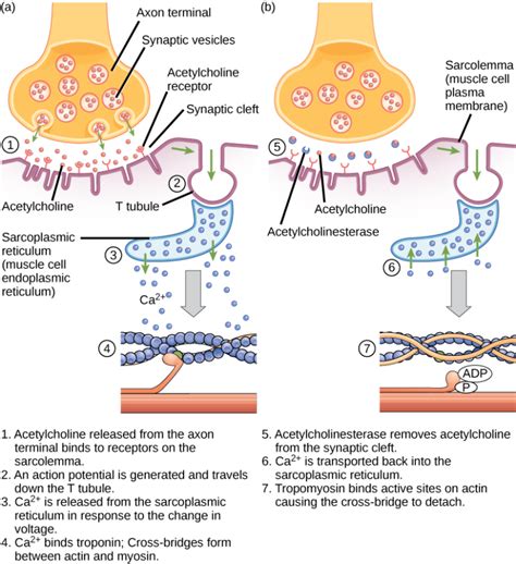
Table of Contents
The Neurotransmitter that Stimulates Skeletal Muscle Contraction: A Deep Dive into Acetylcholine
The human body is a marvel of intricate biological processes, and few are as fundamental as muscle contraction. This seemingly simple action, allowing us to move, breathe, and even think, is orchestrated by a complex interplay of chemical signals and cellular mechanisms. At the heart of this process lies a crucial neurotransmitter: acetylcholine. This article will delve deep into the role of acetylcholine in stimulating skeletal muscle contraction, exploring its synthesis, release, receptor interactions, and the downstream events leading to muscle fiber excitation. We'll also touch upon relevant clinical implications and future research directions.
Understanding the Neuromuscular Junction
Before diving into the intricacies of acetylcholine's role, it's essential to establish the context of the neuromuscular junction (NMJ). The NMJ is the specialized synapse where a motor neuron interacts with a skeletal muscle fiber. This crucial connection facilitates the transmission of nerve impulses, triggering muscle contraction. Imagine it as a highly efficient communication point, ensuring precise control over muscle movement.
The Anatomy of the Neuromuscular Junction
The NMJ is characterized by several key components:
-
Presynaptic terminal (motor neuron axon terminal): This is the site where the motor neuron releases acetylcholine. It contains numerous vesicles packed with acetylcholine molecules.
-
Synaptic cleft: A narrow gap separating the presynaptic terminal from the muscle fiber. This space allows for the diffusion of acetylcholine across the junction.
-
Postsynaptic membrane (muscle fiber membrane, or sarcolemma): This membrane contains acetylcholine receptors, specialized proteins that bind to acetylcholine and initiate the muscle contraction process. The postsynaptic membrane is also highly folded, creating junctional folds that increase the surface area for acetylcholine receptor interaction.
The Acetylcholine Synthesis and Release Cascade
The precise and controlled release of acetylcholine is critical for muscle function. This process involves a series of carefully orchestrated steps:
1. Acetylcholine Synthesis:
Acetylcholine is synthesized within the presynaptic terminal from two precursors:
-
Choline: This is obtained from the diet and is transported into the presynaptic terminal via a sodium-dependent choline transporter.
-
Acetyl CoA: Derived from the mitochondrial metabolism of pyruvate.
The enzyme choline acetyltransferase (ChAT) catalyzes the synthesis of acetylcholine from choline and acetyl CoA. This enzymatic step is crucial; a deficiency in ChAT directly impacts the availability of acetylcholine at the NMJ.
2. Acetylcholine Packaging:
Newly synthesized acetylcholine molecules are then packaged into synaptic vesicles. These vesicles are strategically stored near the presynaptic membrane, ready for release upon stimulation. This packaging ensures a high concentration of acetylcholine at the release site, optimizing transmission efficiency.
3. Acetylcholine Release:
The arrival of a nerve impulse at the presynaptic terminal triggers a cascade of events:
-
Depolarization: The nerve impulse causes depolarization of the presynaptic membrane, opening voltage-gated calcium channels.
-
Calcium Influx: The influx of calcium ions into the presynaptic terminal triggers the fusion of synaptic vesicles with the presynaptic membrane.
-
Exocytosis: This fusion releases acetylcholine into the synaptic cleft via exocytosis. The precise amount of acetylcholine released is tightly regulated and is proportional to the frequency and intensity of the nerve impulse.
Acetylcholine Receptor Activation and Muscle Contraction
Once released into the synaptic cleft, acetylcholine diffuses across the gap and binds to its receptors on the postsynaptic membrane. These receptors are known as nicotinic acetylcholine receptors (nAChRs), named for their sensitivity to nicotine.
Nicotinic Acetylcholine Receptors (nAChRs):
nAChRs are ligand-gated ion channels, meaning they open in response to the binding of a ligand (in this case, acetylcholine). They are composed of five subunits arranged around a central pore. Upon acetylcholine binding, the channel opens, allowing the passage of sodium (Na⁺) and potassium (K⁺) ions.
The End-Plate Potential (EPP):
The influx of sodium ions into the muscle fiber through the open nAChR channels causes a rapid depolarization of the postsynaptic membrane. This depolarization is called the end-plate potential (EPP). The EPP is a graded potential; its amplitude is proportional to the amount of acetylcholine released.
Muscle Fiber Excitation and Contraction:
The EPP initiates a chain reaction leading to muscle fiber contraction:
-
Threshold Potential: If the EPP reaches a sufficient magnitude (threshold potential), it triggers the opening of voltage-gated sodium channels in the adjacent sarcolemma.
-
Action Potential: This leads to the generation of an action potential, a self-propagating wave of depolarization that travels along the muscle fiber membrane.
-
Excitation-Contraction Coupling: The action potential triggers the release of calcium ions from the sarcoplasmic reticulum, the intracellular calcium store within the muscle fiber.
-
Cross-Bridge Cycling: Calcium ions bind to troponin, initiating a conformational change in the troponin-tropomyosin complex. This allows for the interaction of actin and myosin filaments, leading to cross-bridge cycling and muscle contraction.
-
Relaxation: Once the nerve impulse ceases, acetylcholine is rapidly degraded by the enzyme acetylcholinesterase (AChE), located in the synaptic cleft. This hydrolysis of acetylcholine removes it from the receptors, halting further depolarization and allowing muscle relaxation.
Clinical Implications and Diseases
Dysfunction at the neuromuscular junction can lead to several debilitating conditions. Here are some examples:
-
Myasthenia Gravis: An autoimmune disease characterized by the production of antibodies that block or destroy nAChRs. This results in muscle weakness and fatigue.
-
Lambert-Eaton Myasthenic Syndrome (LEMS): A less common autoimmune disorder affecting the presynaptic calcium channels. This reduces acetylcholine release, leading to muscle weakness.
-
Botulism: Caused by the neurotoxin botulinum toxin, which prevents acetylcholine release. This leads to flaccid paralysis.
Understanding the intricate mechanisms of the neuromuscular junction is crucial for the diagnosis and treatment of these and other neuromuscular disorders.
Future Research Directions
Despite significant advancements in our understanding of the neuromuscular junction, several aspects remain areas of active research:
-
Detailed understanding of nAChR subunit composition and function: Different combinations of nAChR subunits can lead to variations in receptor properties and pharmacological responses.
-
Regulation of acetylcholine synthesis, packaging, and release: Exploring the molecular mechanisms underlying the precise control of acetylcholine release is crucial for developing novel therapeutic strategies.
-
Development of novel therapeutic targets for neuromuscular diseases: Research is focused on identifying and targeting specific molecules involved in the pathophysiology of neuromuscular diseases.
Conclusion
Acetylcholine stands as the pivotal neurotransmitter responsible for initiating skeletal muscle contraction. Its journey, from synthesis and packaging to release, receptor activation, and subsequent muscle fiber excitation, is a testament to the elegance and precision of biological processes. Understanding the intricate mechanisms of the neuromuscular junction not only enhances our appreciation of the human body but also provides crucial insights for diagnosing and treating various neuromuscular disorders. Continued research into the nuances of acetylcholine signaling will undoubtedly unveil further intricacies and pave the way for innovative therapeutic approaches in the future. This exploration into the world of acetylcholine is a journey into the fundamental mechanisms that allow us to move and interact with the world around us. The more we learn, the better equipped we are to address the challenges and complexities of neuromuscular function and dysfunction.
Latest Posts
Latest Posts
-
Why Are Most Fossils Found In Sedimentary Rocks
Mar 29, 2025
-
What Is The Gcf Of 45 And 36
Mar 29, 2025
-
What Number Is 45 Of 90
Mar 29, 2025
-
What Is 2 5 As A Decimal
Mar 29, 2025
-
18 As A Percentage Of 60
Mar 29, 2025
Related Post
Thank you for visiting our website which covers about Neurotransmitter That Stimulates Skeletal Muscle Contraction . We hope the information provided has been useful to you. Feel free to contact us if you have any questions or need further assistance. See you next time and don't miss to bookmark.
